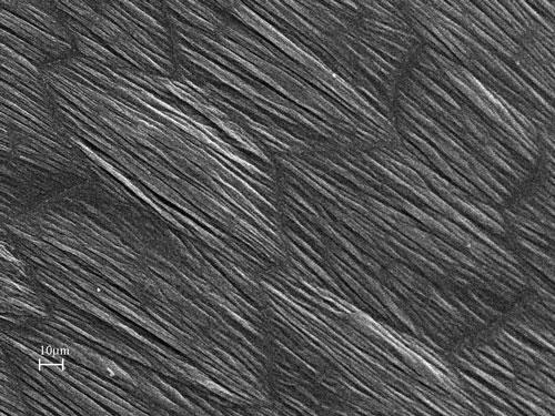
Scanning electron micrograph of the surface of a mouse bladder showing adjacent umbrella cells, tight junctions between them and highly ridged apical membrane due to uroplakin plaques.
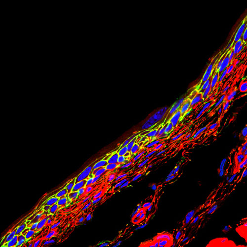
Immunofluorescent staining of a frozen sectioned bladder showing location of aquaporin 3 (green) in basal urothelial cells. Actin is red, nuclei are blue.
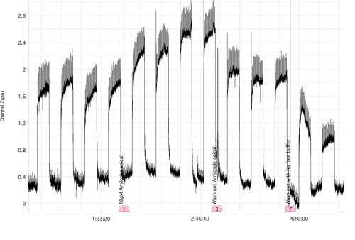
Short circuit currents generated by a mouse bladder mounted in a Ussing chamber and exposed to repeated hydrostatic pressures of 1 cm H2O.
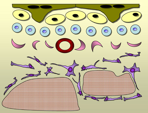
Schematic of bladder showing the diversity of cell types from umbrella cells at top to smooth muscle beneath. Fibroblasts, myofibroblasts and a blood vessel occupy the lamina propria.
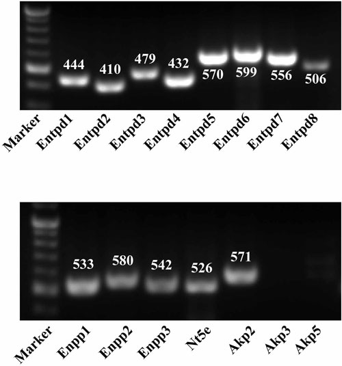
Ectonucleotidase expression in bladder examined by RT-PCR. Taken from Yu et al. (2011) PLoS ONE.
Toluidine blue staining and electron microscopy of the luminal surface of mouse bladder.
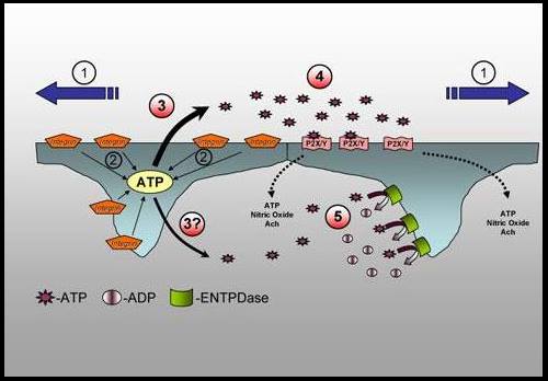
Putative model of integrin mediated signaling by umbrella cells in response to stretch.
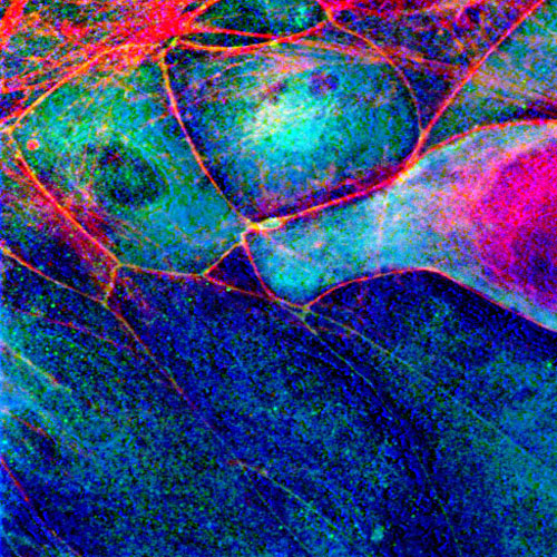
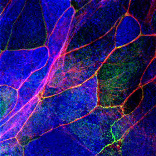
Primary mouse urothelial cells in culture showing cytokeratin (blue), ZO1 at tight junctions (green) and actin (red).
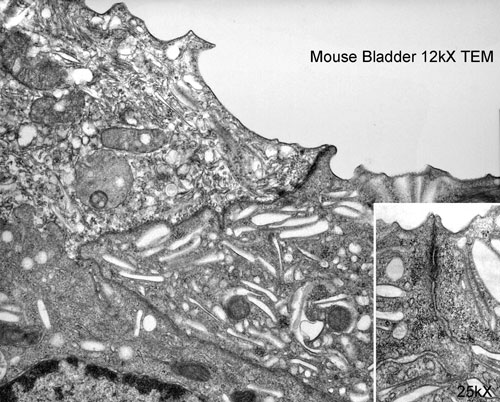
Transmission electron micrograph of umbrella cell apical membrane and tight junction between adjacent cells.
Primary mouse urothelial cells in culture showing uroplakin (blue), ZO1 at tight junctions (green) and actin (red).
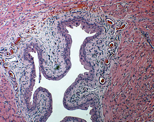
H&E section of mouse bladder.
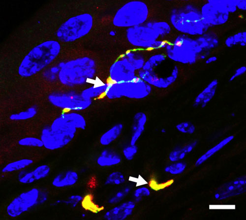
TRPV1(Green) colocalization with neuronal marker, CGRP(Red) within urothelium. Colocalization is indicated by arrows and yellow regions. Taken from Yu et al. Am J Physiol Renal Physiol. 2011 Jan;300:F49.
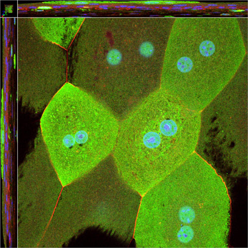
Whole mount confocal microscopy of bladder with GFP-expressing umbrella cells.