Contrast-enhanced Mammography - Another Type of Mammogram
BIDMC Contributor
OCTOBER 29, 2018

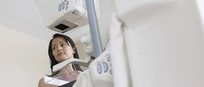
In the fight against breast cancer, there are various diagnostic tools available. One you may not be familiar with is contrast-enhanced mammography (CEM). A CEM is similar to a mammogram in how it's performed with one significant difference — CEM is performed after contrast dye is administered through an IV. The contrast indicates areas of increased blood flow. Because breast cancers generally have more blood vessels, they will often attract the contrast.
For CEM, breast imaging specialists see the findings seen on a conventional mammogram, with the added benefit of seeing areas of abnormal vascularity or blood flow which may be associated with breast cancer.
The imaging portion of the exam takes between 5-10 minutes to complete. Like conventional mammography, CEM is a safe, low-radiation-dose test.
Multiple studies have compared CEM to conventional mammography and have found that CEM is better at finding breast cancer and better at reducing the "false alarms" by more accurately confirming which areas are not cancer. At this time CEM is not a replacement for conventional screening mammography. Rather it is used as a diagnostic tool to evaluate abnormalities questioned on screening mammograms, for patients with breast symptoms and to determine the extent of cancer in patients with known cancer, to name a few.
Sometimes the mammogram and ultrasound aren't able to figure out whether the area is cancer. This is especially true in women with dense breasts, where overlapping breast tissue can make it harder to see a cancer on routine mammograms. This is when the CEM can be helpful. By highlighting areas that take up contrast, CEM can give more information about whether a cancer is present.
CEM can also be used as an alternative to breast MRI since they both use contrast to find breast cancer. CEM can also be used in patients who cannot get MRI due to claustrophobia or having metal in the body.
What's more, CEM is a faster exam than MRI, costs less, and is easier to schedule in a breast imaging department. A radiologist also interprets the CEM while the patient waits in the department, so any extra mammogram or ultrasound images can be performed right away instead of making the patient come back for another visit. Last, the radiologist is able to give the patient her results during the visit, minimizing the anxiety of prolonged waiting.
